When is a Meniscus Injury Not Relevant?
Seeing the diagnosis of “meniscus tear” likely brings many thoughts to mind, along with a variety of different next steps. What do you need to keep in mind when you identify a meniscus lesion during your clinical exam or receive a patient with a meniscus lesion diagnosis?
Arguably, the diagnosis of a meniscus tear always has some relevance – but, how much? Here are several factors to consider when determining the treatment plan for a meniscus injury.
Function
The menisci in the knee, once thought to be dispensable, and routinely removed surgically after derangement, is actually an extremely important stabilizer of the tibiofemoral joint. We now know its absence leads to earlier degeneration of the joint and greater downstream disability as it serves to absorb 50 to 70 percent of compressive forces in the knee!
At the same time, emerging evidence shows that there may be many contributions beyond biomechanical stability that the meniscus brings to joint health. These include biomarkers that can potentially trigger cellular degeneration cascades, and aide or inhibit meniscus repair.
In addition, there are secondary effects of meniscus injury at the cellular level of function for articular cartilage, bone, ligaments, and synovium. Therefore, damage to this structure will never be completely irrelevant.
Outcomes
There are many reports of asymptomatic individuals with meniscus tears. These patients experience no pain or loss of function, despite pathology being identified on imaging exams (MRI, etc). Granted, most of these are in older populations (over 40), and commonly occur in conjunction with knee osteoarthritis (because of the osteoarthritis or in spite of the osteoarthritis? – we still don’t know for sure).
Regardless, the majority of meniscus tears are not amenable to repair (young or old). In the few that are repairable, full healing is still not guaranteed. For example, in one small case series of 11 patients evaluated for healing status after surgery (complete healing, lax healing, scar tissue healing, and failed healing), all patients reported significant clinical improvement, but not one of them fully healed.
In another study, published in the New England Journal of Medicine, surgical outcomes at one year out were found to be no better than physical therapy for patients with a meniscus tear and knee osteoarthritis. “Relevance” should simply lead you to adjust your plan of care based on the presence/absence of a tear, keeping in mind that each patient will be different.
Physical Exam
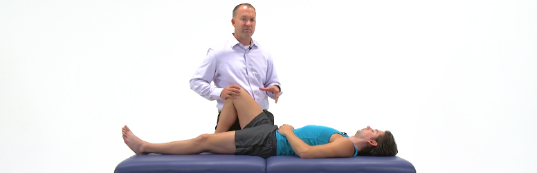
Special tests can help identify the presence of a meniscus tear. However, it can get a little confusing as some studies tout really great diagnostic properties with certain tests, but then systematic reviews (a synthesis of all current studies) come around and point out the poor methodological quality of these studies. They then pool the results, and then all of the sudden the diagnostic properties aren’t as exciting.
The best tests to use may be:
- McMurray’s Test
- Joint Line Tenderness
- Thessaly’s Test
For these tests, it’s important to consider that a recent systematic review showed the sensitivity (the threshold to rule out a tear) and the specificity (the threshold to rule in a tear) to be under 90 percent, and much lower in most cases.
Sometimes, the test will be negative (does not reproduce any pain, catching, clicking, etc), but the gold standard (MRI or arthroscopy) still indicates the presence of a tear. This likely indicates a meniscus tear is stable and may not be as clinically relevant
If you’ve tweaked and twisted and palpated and really tried to make that meniscus squeal…and come up empty-handed, what does that mean? At this point I am less concerned that the MRI might indicate a tear because this clearly does not seem to be the cause of the patient’s symptoms. The diagnosis needs to align with the symptoms and be clinically meaningful.

Subjective Exam
The high rate of asymptomatic meniscus tears requires a proper understanding of the symptoms and a thorough investigation. I would argue that the majority of relevant information will come from the subjective exam.
To start, if you have an MRI report, ensure that the symptoms match with the side of the tear. I’ve treated patients that isolate their symptoms primarily to one side (medial or lateral knee), despite the MRI locating the pathology on the opposite side. What is the disconnect?
Listen for words like “catching” or “locking”, or an explanation of actions that align with these concepts. Sometimes, pain is minimal and the bigger complaint is that the knee locks up when walking. These all may indicate an unstable meniscus, causing it to fold over, slide, or impinge with kinematic movements in the knee.
Take the physical exam a step further to get the complete picture. If you hear that the problem occurs primarily in a loaded position, then make sure you try to reproduce these symptoms in a loaded position (Thessaly versus McMurray’s).
The planning for the objective exam all begins and formulates during the subjective exam. A recent study identified the following examination features that result in a greater likelihood of symptomatic MRI diagnosis in middle-aged patients with knee pain:
- Localized pain, specific to joint line, isolate to side – Identifiable during subjective exam
- Inability to fully bend the knee – Identifiable during subjective exam
- Pain duration
- Lack of varus alignment
- Lack of pes planus
- Absence of joint space narrowing on radiographs.
Make the Features Fit
To completely understand the patient’s symptoms and to form an effective plan of care, you can use the following questions:
- Based on the patient’s history leading up to this consultation, and the nature of their complaint, does it seem likely that the meniscus is causing the problem?
- Do the subjective exam findings align well with the objective exam findings?
- What else could be causing the problem, and how can you go about ruling out those issues beforehand?
- How will your intervention change if you can prove there is a meniscus tear present?
Combining these questions with the functional tests described above is a good way to start to determine if the meniscus tear is relevant or not!
[Click here for references]
- Smith BE, Thacker D, Crewesmith A, Hall M. Special tests for assessing meniscal tears within the knee: a systematic review and meta-analysis. Evid Based Med. 2015 Jun;20(3):88-97.
- Rai MF, McNulty AL. Meniscus Beyond Mechanics: Using Biology to Advance our Understanding of Meniscus Injury and Treatment. Connect Tissue Res. 2017 Mar 29
- Katz JN, Smith SR, Yang HY, Martin SD, Wright J, Donnell-Fink LA, Losina E. Value of History, Physical Examination, and Radiographic Findings in the Diagnosis of Symptomatic Meniscal Tear Among Middle-Aged Subjects With Knee Pain. Arthritis Care Res (Hoboken). 2017 Apr;69(4):484-490
- Deshpande BR, Losina E, Smith SR, Martin SD, Wright RJ, Katz JN. Association of MRI findings and expert diagnosis of symptomatic meniscal tear among middle-aged and older adults with knee pain. BMC Musculoskelet Disord. 2016 Apr 11;17:154.